


HKUST Researchers Develop a Novel Raman Spectroscopy Platform to Characterize Intrinsically Disordered Proteins in Dilute Solution (只供英文版本)
(This article was published on EurekAlert! on April 28, 2021)

It is challenging to analyze proteins at low concentrations, especially for those in a mixture of various conformations such as intrinsically disordered proteins (IDPs). A research team led by Prof. HUANG Jinqing, Assistant Professor of Department of Chemistry at The Hong Kong University of Science and Technology (HKUST), has developed optical tweezers-coupled Raman spectroscopy that can directly probe the structural features of alpha-synuclein, an IDP closely linked to Parkinson’s disease, at the physiological concentration by focusing on individual protein molecules.
IDPs play an important role in biological processes and many of them are associated with incurable neurodegenerative diseases. As a typical IDP, alpha-synuclein lacks a stable 3-D architecture known as secondary structures. It spontaneously undergoes conversions from one secondary structure to another, which could eventually result in the buildup of protein aggregates involved in Parkinson’s disease pathology. However, the transient species during the conversion possess various structures and exist in low population among a dynamic equilibrium mixture. Therefore, their structural features are usually buried under the detection results from traditional measurement techniques, which average the signals detected from large sample quantities and long detection time.
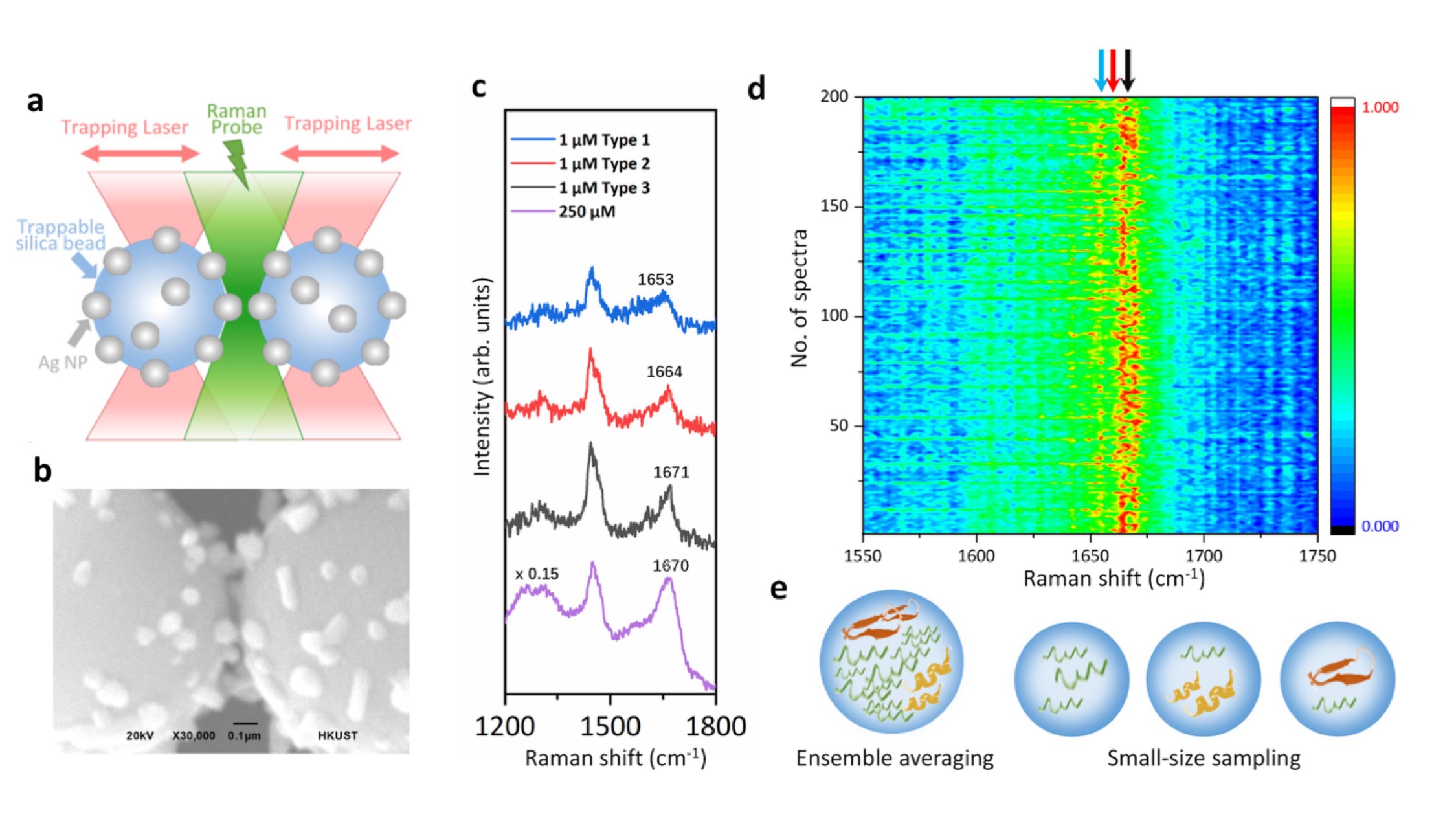
In the study, Prof. Huang and her collaborators integrate optical tweezers and surface-enhanced Raman spectroscopy (SERS) in a novel platform to generate tunable and reproducible SERS enhancements with single-molecule level sensitivity in aqueous environments, in order to characterize these IDPs while maintaining their intrinsic heterogeneity with great biological significance. Specifically, a hotspot can be visualized and controlled by optical tweezers to allow proteins to go through in a microfluidic flow chamber, which makes it convenient to adjust the measurement parameters in the real time for the in situ spectroscopic characterizations. It directly identifies the structural features of the transient species of alpha-synuclein among its predominant monomers at physiological concentration of 1 μM by reducing the ensemble averaging in quantity and in time, providing profound insight to understand the initiation of amyloid protein aggregation. Hence, this SERS platform has great potential to reveal the structural information of IDPs in the dynamic, heterogeneous, and complex biological systems.
“Our strategy enables the precise control of the hotspot between two trapped micrometer-size silver nanoparticle-coated silica beads to improve the SERS efficiency and reproducibility in aqueous detections. Except for the tunable SERS enhancement, the integrated optical tweezers also offer sub-nanometer spatial resolution and sub-piconewton force sensitivity to monitor light-matter interactions in the plasmonic hotspot for extra physical insight. More importantly, our method opens a new door to characterize the transient species of IDPs in dilute solutions, which remains a significant challenge in the biophysics community. Ultimately, it will be exciting to fully exploit the precise force manipulation of the integrated optical tweezers to unfold a single protein inside the controllable hotspot and resolve its structural dynamics from the endogenous molecular vibrations by the integrated Raman spectroscopy.” said, Prof. Huang.
The study was recently published in the scientific journal Nature Communications.
註冊收取我們的最新消息
最新消息
太陽能電池的效能與壽命往往取決於材料之間的微小界面。香港科技大學(科大)研究人員近日參與兩項研究,發現透過精準設計的分子界面層,可顯著提升新一代鈣鈦礦疊層太陽能電池的效能及耐用性。
兩項研究分別刊登於學術期刊《Joule》(影響因子為37.1)及《自然—通訊》,雖然針對不同疊層架構,卻帶出同一個核心訊息:分子界面並非單純連接不同材料的被動層,而是可主動引導鈣鈦礦薄膜結晶、減少能量損失、促進電荷傳輸,並保護器件免受退化影響的重要設計平台。
這兩項研究由科大電子及計算機工程學系助理教授林彥宏教授及科大顯示與光電子全國重點實驗室高級經理楊思恩博士共同帶領,充分結合科大在鈣鈦礦界面設計、光學表徵及疊層器件物理方面的科研優勢。其中,科大電子及計算機工程學系研究助理教授李鳳珠博士領導《Joule》論文的研究工作;科大電子及計算機工程學系博士研究生張青清女士則為《自然—通訊》論文的主要研究團隊成員之一。
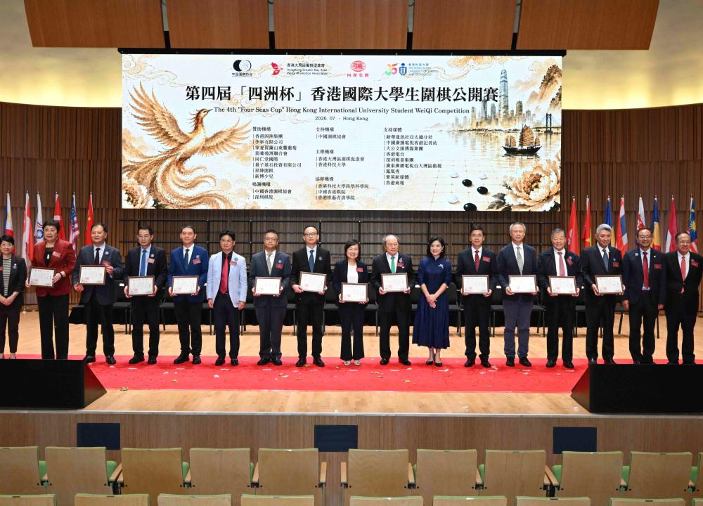
由香港大灣區圍棋促進會及香港科技大學(科大)聯合主辦的第四屆「四洲盃」香港國際大學生圍棋公開賽,於7月14日至18日在科大校園隆重舉行。賽事開幕典禮於今日(7月15日)在科大逸夫演藝中心盛大舉行,並非常榮幸邀請到香港特別行政區政府教育局局長蔡若蓮博士等重量級嘉賓蒞臨主禮。出席開幕儀式的嘉賓亦包括:香港大灣區圍棋促進會會長徐瑩女士、科大副校長(大學拓展)吳宏偉教授、四洲集團創辦人兼主席戴德豐博士、深圳市文化廣電旅遊體育局副局長何建輝先生、中央政府駐港聯絡辦宣傳文體部副部長林枬先生、外交部駐港特派員公署發言人兼新聞及公共關係部主任黃景睿先生、新華社亞太總分社社長孫承斌先生、中央廣播電視總台香港記者站站長王喜凱先生、中央廣播電視總台亞太總站副站長李風先生、先施集團主席、全國政協委員林曉暉先生、香港新聞工作者聯會會長張國良先生及科大跨學科學院院長屈華民教授等。
香港科技大學(科大)一直緊貼時代步伐,因應市場人才需求改革課程。繼去年新推出的生物醫學及健康科學課程成為今年競爭最激烈的課程之一,科大將於新學年推出必修人工智能(AI)通識課程,提升學生在人機協作、AI應用的能力和專業知識。另外,科大的獎學金及學生支援措施與本港主要大學水平相若,並致力透過獎學金、海外學習資助及豐富的發展機會,為學生提供全面而多元的學習體驗。
AI通識課程打造創新人才
為裝備學生迎接人工智能(AI)時代帶來的機遇與挑戰,科大早於2021/22學年率先推出延伸主修(Major + X)課程,讓學生在主修學科以外,選修人工智能、可持續發展等前沿領域,至今已有逾1,450名學生修讀AI相關課程。